Retinal Tear Oct : A case of self-healing retinal pigment epithelium (RPE ... - A retinal tear can occur when the retina pulls away from the outer layers of the eye.
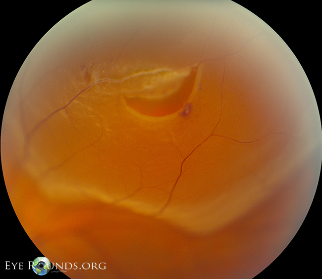
Retinal Tear Oct : A case of self-healing retinal pigment epithelium (RPE ... - A retinal tear can occur when the retina pulls away from the outer layers of the eye.. Typically, however, the vitreous separates without any ill effects on the retina. Several months later, cynthia said it seems as if she is looking through a snow globe. Learn what to expect from a dilated eye exam. The ring of pigmentation (blue arrows) is a reactive repair due to separation of neurosensory retina and the retinal pigment epithelium. The macula is a pigmented area at the centre of the retina.
4 octa may or may not show any significant microvascular changes in the superficial or deep capillary plexuses. It is one of the most common ocular emergencies today, most frequently affecting the middle aged and elderly. Oct raster scan reveals a subclinical retinal tear with minimal surrounding subretinal fluid in a patient with acute onset vitreous hemorrhage and proliferative dr. If you suspect that it's happened to you, you should consult an optician for a comprehensive eye exam as soon as possible. Chiang a, chang lk, yu f, sarraf d.
Peripheral Retinal Degenerations as a Risk Factor for ... from i2.wp.com Flashes of light (photopsia) are another common symptom. However, occasionally, the flap may be pulled free (avulsed) and be seen as an elongated operculum floating above the break. (c) a large atrophic hole noted in routine examination and subsequently treated with laser. The retina plays a vital role in vision. Because of the severity of this condition, it is an excellent topic for medical students to review. Chiang a, chang lk, yu f, sarraf d. Oct images can be used to diagnose many retina related eyes diseases. A retinal tear can occur when the retina pulls away from the outer layers of the eye.
This can have the appearance of someone shaking pepper in your vision.
Oct is heavily used by ophthalmologists to obtain high resolution images of the eye retina. Typically, however, the vitreous separates without any ill effects on the retina. If you suspect that it's happened to you, you should consult an optician for a comprehensive eye exam as soon as possible. Chan ck, meyer ch, gross jg, abraham p, nuthi as, kokame gt et al. In addition, if the laser is coded as 67145, and happens today or tomorrow, modifier 57 would also be needed in addition to modifier 24. Retinal detachment is an important cause of decreased visual acuity and blindness. The laser sealed the tear in cynthia's retina, but the afternoon after her treatment, another black octopus streaked across her vision. Optical coherence tomography (oct) signs include increased reflectivity and thickness of ellipsoid zone (ez) with disruption of interdigitation zone (iz) and occasionally inner retinal layers. Although a tear can usually be repaired successfully, it is considered a serious condition. Both of these tests are painless. Determine macular status in retinal detachment (b) oct findings after 1 day in an eye with surgery using dispersive ovd (arrows) (dispersive ovd group). The doctor may press on your eyelids to check for retinal tears, which may be uncomfortable for some people.
In addition, if the laser is coded as 67145, and happens today or tomorrow, modifier 57 would also be needed in addition to modifier 24. Optical coherence tomography (oct) findings after vitrectomy with application of dispersive ophthalmic viscosurgical device (ovd). A retinal detachment is usually diagnosed clinically and with exam, but shallow macular detachments are sometimes hard to appreciate early on. Although a tear can usually be repaired successfully, it is considered a serious condition. Yes, because the care for the retinal tear eye is unrelated to the postoperative care for the slt.
Rhegmatogenous retinal detachment: The University of Iowa ... from webeye.ophth.uiowa.edu Flap or horseshoe retinal tear a flap (horseshoe) tear results from vitreous traction that pulls a tear of sensory retina that almost always remains attached at the anterior margin of the break. However oct demonstrates crystalline deposition within the inner retinal layers. However, occasionally, the flap may be pulled free (avulsed) and be seen as an elongated operculum floating above the break. Back she went to the eye doctor, who discovered that when the tear occurred it nicked a blood vessel. A retinal tear can occur when the retina pulls away from the outer layers of the eye. It is when the retina has a tear or hole, like a rip in cloth. Both of these tests are painless. Several months later, cynthia said it seems as if she is looking through a snow globe.
Fa, erg and color vision are typically normal;
A torn retina is a serious problem that makes your vision blurry. On oct, rpe tears appear as hyperreflective tissue rolled up underneath the neurosensory retina, with a free edge of wavy rpe at the margins of an area with exposed bruch's membrane and absent overlying rpe (fig. This is known as a rhegmatogenous retinal detachment. It is when the retina has a tear or hole, like a rip in cloth. 4 octa may or may not show any significant microvascular changes in the superficial or deep capillary plexuses. Chan ck, meyer ch, gross jg, abraham p, nuthi as, kokame gt et al. Oct images can be used to diagnose many retina related eyes diseases. A retinal tear can allow the liquid part of the vitreous to escape behind the retina and separate the retina from its underlying attachments (and blood supply). A torn retina often leads to a more serious condition called a detached retina.this is where the retina is lifted away from the back of the eye. Fa, erg and color vision are typically normal; The laser sealed the tear in cynthia's retina, but the afternoon after her treatment, another black octopus streaked across her vision. The macula is a pigmented area at the centre of the retina. Because of the severity of this condition, it is an excellent topic for medical students to review.
The doctor may press on your eyelids to check for retinal tears, which may be uncomfortable for some people. Flap or horseshoe retinal tear a flap (horseshoe) tear results from vitreous traction that pulls a tear of sensory retina that almost always remains attached at the anterior margin of the break. Posterior vitreous detachment posterior vitreous detachment(pvd) occurs as a natural aging progress but can lead to the risk for severe visual impairment if associated with retinal tears that can then develop into a retinal detachment. A retinal detachment is usually diagnosed clinically and with exam, but shallow macular detachments are sometimes hard to appreciate early on. Chan ck, meyer ch, gross jg, abraham p, nuthi as, kokame gt et al.
(PDF) Spontaneous Large Serous Retinal Pigment Epithelial Tear from www.researchgate.net Fa, erg and color vision are typically normal; (c) a large atrophic hole noted in routine examination and subsequently treated with laser. On oct, rpe tears appear as hyperreflective tissue rolled up underneath the neurosensory retina, with a free edge of wavy rpe at the margins of an area with exposed bruch's membrane and absent overlying rpe (fig. The areas where the retina detaches lose their blood supply and stop working, causing you to lose vision. Although a tear can usually be repaired successfully, it is considered a serious condition. Oct is heavily used by ophthalmologists to obtain high resolution images of the eye retina. However, occasionally, the flap may be pulled free (avulsed) and be seen as an elongated operculum floating above the break. If you suspect that it's happened to you, you should consult an optician for a comprehensive eye exam as soon as possible.
(a) oct findings on day 7 in the control group.the retinal tear is open and curled, and retinal detachment persists;
Determine macular status in retinal detachment Learn what to expect from a dilated eye exam. The macula is a pigmented area at the centre of the retina. If your eye doctor still needs more information after a dilated eye exam, you may get an ultrasound or an optical coherence tomography (oct) scan of your eye. Yes, because the care for the retinal tear eye is unrelated to the postoperative care for the slt. In addition, if the laser is coded as 67145, and happens today or tomorrow, modifier 57 would also be needed in addition to modifier 24. (b) oct findings after 1 day in an eye with surgery using dispersive ovd (arrows) (dispersive ovd group). It is one of the most common ocular emergencies today, most frequently affecting the middle aged and elderly. Chiang a, chang lk, yu f, sarraf d. If any doubt, a retinal oct can demonstrate a detachment easily. Retinal tears when a retinal tear or hole hasn't yet progressed to detachment, your eye surgeon may suggest one of the following procedures to prevent retinal detachment and preserve vision. Damage to the retina can cause vision loss and even permanent blindness. Atrophic retinal hole (red arrows) noted both on (a) fundus photograph and (b) oct.
Because of the severity of this condition, it is an excellent topic for medical students to review retinal tear. (c) a large atrophic hole noted in routine examination and subsequently treated with laser.


0 Komentar